
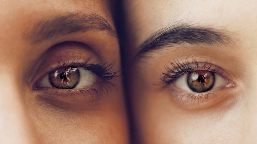
The human eye – a mystery to most of us.
How much do we really know about our eyes? Have you ever wondered how they work, or how they’re able to transmit images to the brain to allow us to see? In this article, we explore all the parts that make up the eye and shed some light on how it all works.
Parts of the human eye
The human eye is a sensory organ, housed in the cavity known as the orbit, surrounded by skin, muscles, and a thick layer of tissue. Many parts of the eye work together to allow it to function properly and, when well cared for, to protect it from certain sight-threatening diseases and injuries.
Sclera
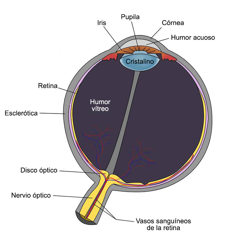
The sclera is the white part of the eye – a fibrous layer made of collagen that borders the cornea.
Function of the sclera
The sclera surrounds most of the eye, front and back, offering protection by preserving its structure and shielding it from injury.
Cornea
The cornea is the transparent, dome-shaped part that covers the iris and pupil externally, behind a thin tear film.
Function of the cornea
The primary function of the cornea is vision. As one of the first areas reached by light as it enters the eye, the cornea is responsible for a large portion of our focusing power. If the cornea is too flat, too curved, or has an irregular shape, it causes refractive errors such as short-sightedness, long-sightedness, or astigmatism.
Iris
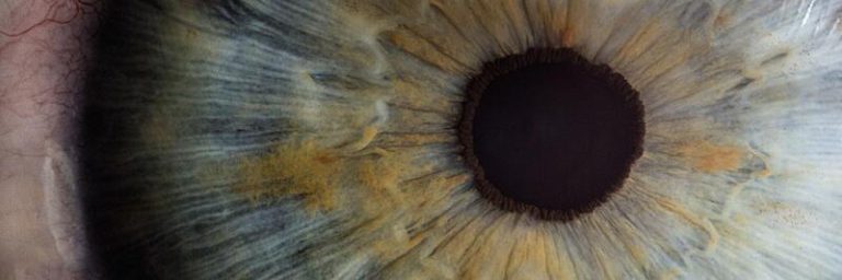
The iris is the brown, blue, or green part of our eye. Located behind the cornea but in front of the lens, the iris surrounds the opening known as the pupil.
Function of the iris
The iris regulates light, allowing the right amount to pass through the pupil. It dilates in low light and contracts in bright environments or when focusing on nearby objects.
Lens
The lens is the transparent tissue located directly behind the pupil.
Function of the lens
When we are born, the lens is flexible and reshapes to allow the eye to focus at different distances. As we age, the lens loses its elasticity, leading to an inevitable age-related condition called presbyopia.
Vitreous body
The vitreous body is a gel-like substance found behind the lens in the back of the eye. It is made up of a combination of water, collagen, and proteins.
Function of the vitreous body
The vitreous body plays a key role in helping the eye maintain its spherical shape, as well as protecting it from trauma and injury. It occupies two thirds of the eye’s structure and is attached to the retina.
Retina
The retina is a light-sensitive tissue that lines the back of the eye.
Function of the retina
The retina acts as a light receptor. Like the film in a camera, it processes light through photoreceptor cells to detect colour and detail, helping the optic nerve to send impulses to the brain and convert them into images.
Optic nerve
The optic nerve is the largest nerve in the eye. It is located at the back of the eye and connects to the brain.
Function of the optic nerve
The optic nerve sends signals and impulses to the visual cortex of the brain, which is responsible for our vision. These impulses are transmitted through millions of optic nerve fibres.
How do eyes work?
All the structures mentioned above must work together for us to perceive vision. If any of these parts are not functioning correctly – due to eye conditions, diseases, or injuries – our vision will be affected.
Sight
The first step towards vision occurs when light passes through the thin tear film and enters the cornea, where the eye begins to focus. After the cornea, light passes through the pupil (the opening of the iris), which controls the amount of incoming light.
The lens and cornea work together to allow light to be correctly focused as it passes through the vitreous body and onto the retina.
The retina transmits the received image to the brain via the optic nerve. Our brain then processes the information and constructs the image we see – our vision.
Colour perception
Most people see the world in colour, and they have the retina to thank for it.
The photoreceptors in the retina contain two types of sensory cells: rods and cones. Rods handle changes in brightness and vision in low-light environments, while cones are responsible for colour processing.
Each type of cone is sensitive to light at different wavelengths. Blue colours react to shorter wavelengths, greens to medium wavelengths, and reds to longer wavelengths.
The cones detect the wavelength of each colour and send this information to the brain.
Parts susceptible to conditions that can affect vision
From the front to the back of the eye, every part must work together to provide clear vision. However, the eye is also a sensitive organ and, if not properly cared for, can be susceptible to a range of conditions.
Cornea
As the outermost part of the eye, the cornea is most exposed to external hazards and surface diseases.
Injuries and abrasions can cause corneal scarring and distorted vision.
Allergies to pollen, pets, or dust can irritate the outer part of the eye, causing watering, itching, or redness.
The cornea can also be affected by conditions such as keratoconus – a disease that causes the cornea to thin and change shape, leading to increasingly blurry and distorted vision.
Retina
The retina lines the back of the eye and is therefore protected from most external factors, but it remains susceptible to damage.
Retinal diseases can be caused by a range of factors, including age, trauma, general health conditions, or genetics.
The most common retinal disorder is diabetic retinopathy, caused by high blood sugar levels in people with type 1 or type 2 diabetes.
Macular degeneration is another common eye condition that affects the macula (the centre of the retina). It is usually age-related and affects the central visual field.